Brain Organoids: Miniature Neural Tissue and the Questions It Raises
Scientists grow simplified clusters of human neural tissue from stem cells to study brain development. These organoids provide biological insights while raising difficult questions regarding the future potential for sensory perception and consciousness.
Key points
- Organoids form through self-organization as pluripotent stem cells differentiate into specific neural lineages, mimicking the basic structural layers found in a developing human foetal brain.
- Current models lack blood vessels and immune cells, limiting their growth to a few millimetres and preventing the complexity required for mature cognitive function or meaningful thought.
- Researchers use these tissues to observe how genetic mutations affect synaptic pruning and cellular migration, offering a dynamic view of developmental disorders that animal models cannot provide.
- Ethical concerns centre on the eventual integration of organoids into robotics or silicon systems, necessitating a framework for identifying indicators of nascent sentience in biological hardware.
Modern neurobiology faces a persistent constraint in the physical opacity of the living human brain. While animal models offer valuable insights into basic neural functions, the unique genetic and developmental trajectory of the human neocortex remains difficult to observe in real time. Post-mortem tissue represents a fixed, terminal state, and non-invasive imaging lacks the resolution to track the movement of individual cells or the formation of specific synaptic junctions.
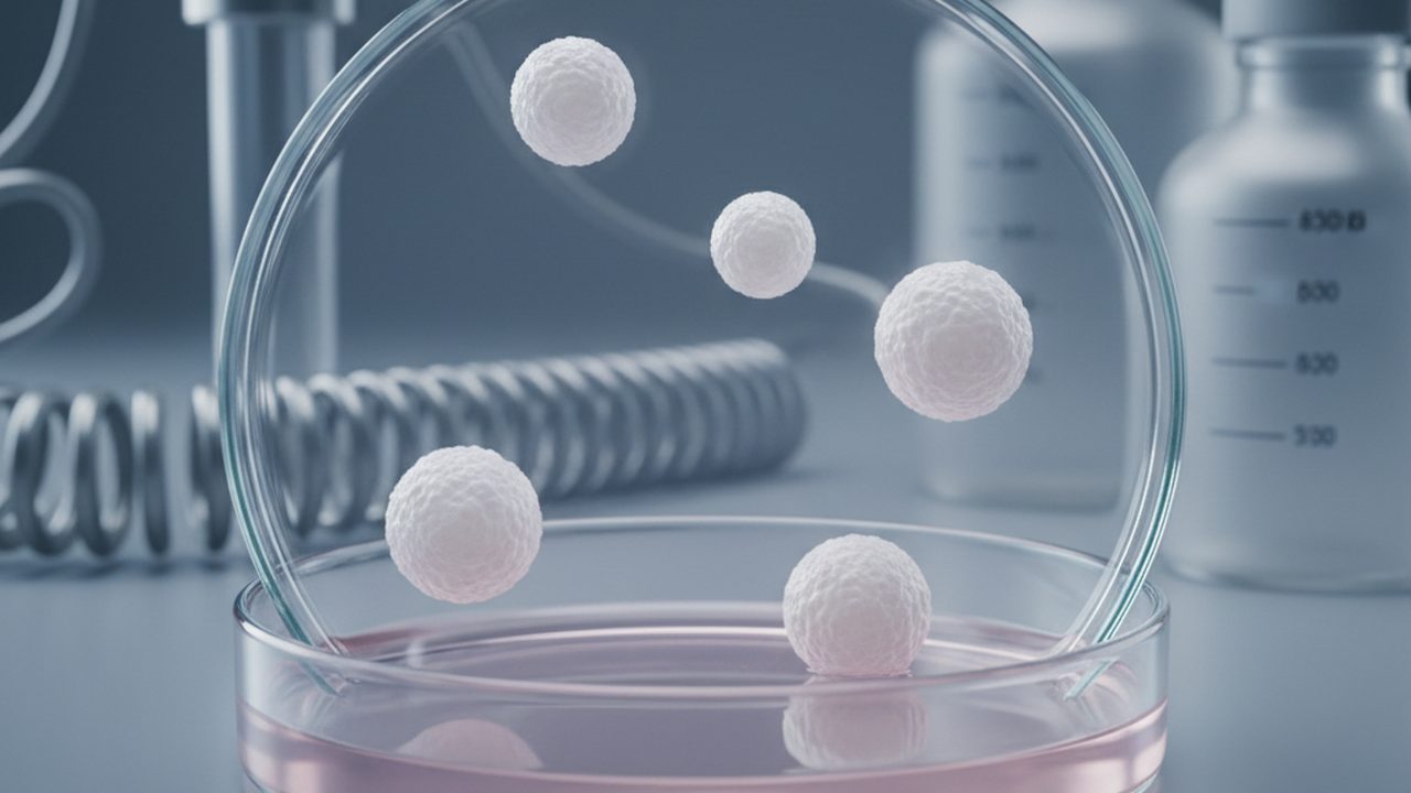
To address this, researchers have turned to three-dimensional neural cultures, commonly known as brain organoids. These are clusters of human cells grown in laboratories that replicate certain structural and functional aspects of the developing brain. They are not miniature brains in a functional sense, as they lack the sensory input, vascular systems, and bodily integration required for consciousness or cognition. Instead, they serve as simplified models for studying how the human brain builds itself from the ground up.
The utility of an organoid lies in its capacity to mimic the early stages of embryonic development. By directing the growth of stem cells into neural lineages, scientists can observe the mechanics of cell division, migration, and differentiation in a controlled environment. This allows for the study of neurodevelopmental disorders, such as microcephaly or lissencephaly, by comparing organoids grown from patient-derived cells against those from healthy controls.
Despite their utility, organoids are limited by their environment. In the absence of a circulatory system to deliver nutrients and remove waste, the interior of a maturing organoid often suffers from necrosis. This physical limitation caps their size at a few millimetres and prevents them from reaching the complexity of a late-stage foetal brain. The field currently exists in a state of cautious exploration, balancing the technical potential of these tissues with a growing need to define the ethical boundaries of creating human neural material in vitro.
The cellular origin of neural organoids
The creation of a brain organoid begins with pluripotency. Pluripotent stem cells possess the capacity to differentiate into any cell type in the human body, provided they receive the correct biochemical cues. In contemporary research, these cells are typically induced pluripotent stem cells, derived from adult skin or blood cells that have been genetically reprogrammed to an embryonic-like state. This allows researchers to study a specific individual’s genetic profile without the use of embryonic tissue.
To initiate the transition into neural tissue, these stem cells are placed into a suspension culture where they aggregate into small spheres called embryoid bodies. Researchers then apply a series of growth factors that inhibit other developmental paths, such as those leading to muscle or bone, and promote the ectodermal lineage. The ectoderm is the outermost layer of the early embryo, which eventually gives rise to the skin and the nervous system.
Once the cells are committed to a neural identity, they are often embedded in a scaffold of extracellular matrix, usually a protein-rich gel that provides structural support. This gel mimics the physical environment of the developing embryo, allowing the cells to expand outwards rather than collapsing into a flat layer. Within this environment, the cells begin to respond to internal signalling molecules, initiating a process of specialisation that mirrors the earliest weeks of human gestation.
Principles of biological self-organisation
A defining characteristic of organoids is that they are not assembled by hand; they self-organise. This process relies on the innate ability of cells to interpret their surroundings and communicate with their neighbours. Even in a homogeneous cluster of cells, slight variations in the concentration of signalling proteins create gradients. Cells detect these gradients and change their behaviour accordingly, a phenomenon known as morphogenesis.
The primary mechanism driving this organisation is differential adhesion. Different types of neural cells express different proteins on their surfaces, which act like biological Velcro. Cells with similar surface proteins tend to stick together more tightly than those with different ones. As the cells divide and mature, they naturally sort themselves into distinct layers and clusters. This occurs without an external blueprint, guided only by the laws of physics and the genetic instructions within each cell.
This self-organisation results in the formation of neural rosettes, which are circular arrangements of cells that mimic the structure of the neural tube. These rosettes serve as the functional units of the organoid, acting as the sites where new neurons are generated. The spontaneous emergence of these structures demonstrates that the basic architectural plan of the human brain is encoded within the cells themselves, rather than being entirely dependent on the complex environment of the womb.
The capacity for stem cells to spontaneously form complex, layered structures suggests that the fundamental blueprint of the brain is intrinsic to the cells.
Observing the architecture of the developing cortex
The human neocortex is structured into six distinct horizontal layers, each containing specific types of neurons that perform different computational tasks. In a developing embryo, this architecture is formed through the birth and migration of cells. Radial glial cells act as both progenitors and physical tracks, stretching from the inner ventricular zone to the outer surface of the brain. New neurons climb these glial fibres to reach their final destination, with later-born neurons migrating past their predecessors to form the upper layers.
Organoids allow researchers to watch this process in a way that was previously impossible. By using fluorescent markers to label different cell types, scientists can track the birth of excitatory neurons and their subsequent migration. They have observed that organoids can replicate the "inside-out" pattern of cortical development, where the deeper layers of the cortex form before the superficial ones. This suggests that the timing of cell birth is a highly conserved biological programme.
However, the architecture in an organoid is often disordered compared to a natural brain. Instead of a single, continuous sheet of cortex, an organoid may contain multiple small, folded regions of cortical tissue facing in different directions. While the local layering within these regions can be remarkably accurate, the global topography is fragmented. This makes organoids excellent for studying how individual neurons find their place, but less effective for studying how different regions of the brain, such as the visual and motor cortices, connect to one another.
Comparison between organoids and whole-organ biology
While the term brain organoid suggests a miniature organ, the reality is more akin to a collection of specific tissues. A human brain is a vascularised organ integrated with a central nervous system, receiving constant sensory feedback and hormonal regulation. An organoid, by contrast, is an isolated biological system. It lacks microglia—the brain's immune cells—unless they are specifically added, and it lacks the blood vessels necessary to sustain large-scale growth.
The absence of vasculature is the most significant divergence from whole-organ biology. In a living brain, oxygen and glucose are delivered through a dense network of capillaries. In an organoid, these nutrients must diffuse from the surrounding liquid medium into the centre of the tissue. As the organoid grows, the distance to the centre becomes too great for diffusion to be effective, leading to a core of dead cells. This limits the maturity of the tissue, as it cannot reach the developmental stages where complex neural circuits and long-range connections are fully established.
Furthermore, the lack of sensory input means that the electrical activity observed in organoids is fundamentally different from that of a functioning brain. While researchers have detected spontaneous bursts of electrical activity and even basic forms of synchrony, these signals do not represent thoughts or perceptions. They are the physiological equivalent of a motor idling. Without the feedback loops provided by eyes, ears, and skin, the neural circuits in an organoid cannot be calibrated or refined through experience. Consequently, while organoids are powerful models for early development and cellular pathology, they remain simplified abstractions of the human brain.
The barrier of oxygen and nutrient flow
The most significant physical constraint on the growth of brain organoids is the absence of a vascular system. In a living organism, a dense network of capillaries ensures that no cell is more than a few microns away from a blood vessel. This infrastructure delivers oxygen and glucose while simultaneously removing metabolic waste products like carbon dioxide and lactic acid. Because laboratory-grown organoids lack these vessels, they rely entirely on passive diffusion. Molecules must travel from the surrounding nutrient broth into the depths of the tissue by moving down a concentration gradient.
As an organoid grows beyond a diameter of roughly two or three millimetres, this passive transport becomes insufficient. The cells at the outer surface consume the available oxygen, leaving the interior cells to suffocate and die. This process creates a necrotic core, a pocket of dead tissue that limits the overall health and longevity of the culture. To mitigate this, researchers have experimented with mechanical stirring and rotating bioreactors, which keep the medium in constant motion to maximise exposure. Some teams have even attempted to co-culture neural cells with endothelial cells, which form the lining of blood vessels, in the hope that they will self-organise into a plumbing system.
While these methods have extended the lifespan of organoids, they have not yet replicated the high-pressure, directed flow of a heart-driven circulatory system. Without this, the tissue cannot develop the thick, multi-layered cortex characteristic of a human brain. The resulting structures remain analogous to the brain of a first-trimester human embryo. This structural ceiling means that while organoids can show us how individual neurons sprout and migrate, they cannot yet model the large-scale architectural folding or the precise layering of the adult cerebral cortex.
Modelling genetic and neurodevelopmental disorders
Despite their structural limitations, organoids have become essential for studying how genetic variations alter the early stages of brain assembly. Traditionally, researchers relied on animal models, particularly mice. However, the human brain undergoes developmental processes, such as the massive expansion of the outer subventricular zone, which are absent in rodents. By using induced pluripotent stem cells (iPSCs) derived from patients with specific conditions, scientists can grow organoids that carry the exact genetic code of the individual.
This approach has been particularly effective in studying microcephaly, a condition where the brain is significantly smaller than average. In these organoids, researchers have observed that neural stem cells prematurely transition into mature neurons. This exhausts the pool of progenitor cells too early, resulting in a smaller total number of cells and a reduced tissue volume. Because this process is visible in the laboratory dish, researchers can test chemical compounds to see if they can slow this premature differentiation.
The same principle applies to complex neuropsychiatric conditions. For instance, in models of autism spectrum disorder, organoids have revealed imbalances between excitatory and inhibitory neurons. By observing the way these cells migrate and connect, it is possible to identify whether a specific mutation affects the speed of cell movement or the way synapses are formed. This creates a platform for high-throughput drug screening. Rather than testing a drug on a patient, researchers can apply it to hundreds of identical organoid samples to measure the molecular response. This moves the field closer to a model of personalised medicine, where a patient’s own cells provide the blueprint for their treatment.
The ability to observe human-specific developmental pathologies in a controlled environment bypasses many of the biological discrepancies found in animal models.
The shift toward biological computing
A new direction in the field involves integrating brain organoids with silicon technology. This area, often termed organoid intelligence or biological computing, seeks to use the natural processing power of neural tissue to perform computational tasks. Neurons are inherently more energy-efficient than traditional silicon chips when it comes to pattern recognition and learning. While a supercomputer requires megawatts of power to simulate a small fraction of a brain, the human brain operates on roughly twenty watts.
In these experimental setups, organoids are grown on microelectrode arrays. These arrays allow researchers to both record the electrical pulses produced by the neurons and stimulate the tissue with specific electrical patterns. By providing a feedback loop, researchers can train the organoid to respond to inputs. In one notable demonstration, a mass of neural cells was taught to perform a simple task in a simulated environment by receiving electrical cues when it made a correct or incorrect move.
However, the mechanism behind this is not "thought" in any human sense. It is a manifestation of synaptic plasticity, the process by which neural connections strengthen or weaken based on activity. The organoid is not aware of the task; it is simply physically adapting its circuitry to the electrical stimuli it receives. The long-term goal for some engineers is to create hybrid systems where biological tissue handles complex, non-linear processing while silicon handles stable memory storage and high-speed arithmetic. This remains a speculative engineering goal, as maintaining the viability of biological tissue alongside electronic hardware for long periods is technically demanding.
Defining the boundaries of sentience
The transition from a passive clump of cells to an active computational unit raises questions about the point at which neural tissue might be said to possess sentience. In the context of bioethics, sentience is usually defined as the capacity to have subjective experiences, such as the ability to feel pain or pleasure. Consciousness is a higher bar, involving a sense of self and the integration of diverse sensory streams into a single narrative.
Currently, there is a broad consensus that brain organoids do not possess either. They lack the necessary hardware for consciousness, such as a thalamus to relay sensory information or a sufficiently complex cortical structure to integrate it. The electrical activity recorded from them is disorganised and lacks the information density seen in even the simplest living brains. However, as techniques for vascularisation and sensory integration improve, the gap between an organoid and a living brain may narrow.
The difficulty lies in the fact that we lack a definitive physical marker for the emergence of consciousness. If an organoid were to show complex, rhythmic patterns of activity similar to those found in a conscious human, it would be unclear if the tissue is actually "experiencing" anything or if it is merely a sophisticated simulation. Some philosophers argue that without an internal drive for survival and a body to protect, a brain in a dish can never truly be sentient. Others suggest that we should apply the principle of precaution, treating any tissue that shows advanced neural synchrony with a degree of moral consideration.
A framework for ethical governance
As the technology advances, the governance of organoid research must move beyond standard laboratory safety protocols. There are several distinct areas where ethical standards are currently being debated and refined.
- The provenance of donor material: When individuals donate skin or blood cells for stem cell research, they often do so under a general consent framework. It is unclear if specific consent should be required when those cells are used to create neural tissue that could potentially be kept alive for years or integrated into computers.
- The status of human-animal chimeras: To overcome the lack of a vascular system, some researchers transplant human organoids into the brains of living rats or mice. The organoid integrates with the host’s blood supply and neural circuitry. This raises questions about whether the cognitive abilities of the host animal are altered and what the moral status of such an animal should be.
- The definition of "death" for lab-grown tissue: If an organoid were to reach a level of complexity where it demonstrated sentient-like activity, there would need to be protocols for how and when such a culture could be terminated.
Currently, oversight is managed by institutional review boards that evaluate research on a case-by-case basis. There are no international laws specifically governing the complexity of brain organoids. Proposals for future governance include the creation of a "neural complexity scale" to categorise organoids based on their developmental stage and electrical activity. Reaching certain thresholds on this scale would trigger more stringent ethical reviews and limitations on what could be done with the tissue.
The current state of the science
At present, brain organoids are firmly established as a valuable tool for molecular biology. Their ability to replicate the early stages of human neurogenesis has provided insights that were previously impossible to obtain. They have reliably demonstrated the mechanisms behind several genetic disorders and provided a platform for testing the toxicity of new drugs on human cells.
What is contested is the extent to which these models can ever truly represent the mature human mind. The physical limitations of oxygen transport and the absence of sensory input remain significant hurdles. Furthermore, the question of whether a detached mass of neurons can ever achieve a state of consciousness remains a subject of intense philosophical and scientific debate, with no clear consensus on how such a state would even be measured.
The picture would change significantly if a reliable method for full vascularisation were developed, or if an organoid were successfully integrated with a functional sensory system, such as a laboratory-grown retina. Such a breakthrough would move the field from the study of isolated cells to the study of integrated neural systems, bringing the theoretical questions of sentience and moral status into much sharper relief. For now, the organoid remains a simplified map of a vastly more complex territory.