Deep Brain Stimulation: Neurology's Most Successful Implant
Deep brain stimulation uses high-frequency electrical pulses to stabilise neural circuits. This feature examines the shift from constant stimulation to adaptive systems that respond to real-time neurological signals.
Key points
- Electrodes implanted in the subthalamic nucleus or globus pallidus use electrical interference to override the erratic firing patterns characteristic of late-stage Parkinson's disease.
- The therapy operates on a circuit level rather than a chemical one, offering a mechanical alternative when pharmacological treatments like levodopa become ineffective or cause dyskinesia.
- Closed-loop systems represent a significant advancement by sensing local field potentials, allowing the device to deliver stimulation only when specific pathological rhythms are detected.
- Emerging research into neurotropic targets suggests that stimulation may improve psychiatric conditions by modulating the connectivity between the prefrontal cortex and the limbic system.
For most of the twentieth century, clinical neurology treated the brain as a chemical broth. The primary method for correcting tremors, rigid limbs, or persistent low mood was the introduction of synthetic molecules into the bloodstream. These compounds were intended to dock with specific receptors across the entire organ, shifting the balance of neurotransmitters like dopamine or serotonin. While this systemic approach remains the standard of care for many conditions, its lack of spatial precision introduces a trade-off. A drug intended to quiet a motor tremor in one region of the brain may simultaneously cause hallucinations or metabolic dysfunction by interacting with identical receptors in another.
The limitations of pharmacology led to the development of deep brain stimulation, or DBS. Rather than attempting to bathe the brain in a corrective chemical, DBS treats the organ as a network of electrical circuits. It operates on the principle that many neurological and psychiatric conditions are not merely global chemical imbalances, but are instead failures of specific signalling loops. When these circuits begin to fire with excessive synchrony or at inappropriate frequencies, the resulting symptoms manifest as the physical stuttering of Parkinson’s disease or the obsessive cycles of severe depression.
Physicians and engineers have found that by inserting a thin, insulated wire into a precise anatomical coordinate and applying a high-frequency current, they can interrupt these pathological signals. The effect is often immediate. A patient whose limbs were frozen by rigidity may regain fluidity of movement the moment the device is activated. This intervention does not heal the underlying tissue, nor does it replace the lost neurons. Instead, it acts as a functional pacemaker for the brain, overriding erratic internal signals with a steady, external rhythm.
This technology has transitioned from an experimental last resort to a standard intervention for movement disorders. More recently, its application has broadened to include treatment-resistant depression and obsessive-compulsive disorder. The success of the procedure has forced a shift in how researchers conceptualise mental illness, moving away from vague notions of personality or temperament toward a concrete understanding of circuit-based dysfunction.
The mechanical transition from chemistry to electricity
The shift from chemical to electrical intervention reflects a fundamental property of the neuron. While communication between neurons occurs via the release of neurotransmitters across a synapse, the transmission of information within a single neuron is electrical. An action potential, or nerve impulse, travels along the axon as a wave of depolarisation. Deep brain stimulation bypasses the chemical signaling step by using an external electric field to trigger these action potentials directly.
In a pharmacological model, a patient with Parkinson’s disease might take levodopa to compensate for a lack of dopamine. This chemical is meant to facilitate the smooth flow of information through the basal ganglia, a cluster of structures deep within the brain responsible for coordinating movement. However, as the disease progresses, the brain’s ability to process these chemicals becomes volatile. Patients often cycle between periods of mobility and periods of uncontrolled, jerky movements known as dyskinesia. The chemical supply is simply too blunt a tool to match the brain’s millisecond-by-millisecond requirements.
Electrical stimulation offers a different level of control. By placing an electrode directly into the dysfunctional circuit, clinicians can modulate the activity of thousands of neurons simultaneously. This is not a natural signal, but it is a predictable one. The electrical field creates a zone of influence where the erratic, bursting patterns of a diseased brain are suppressed and replaced by a constant frequency. This transition from chemistry to electricity allows for a continuous, adjustable therapy that does not rely on the digestive system or the blood-brain barrier for delivery.
Anatomy of the electrode and pulse generator
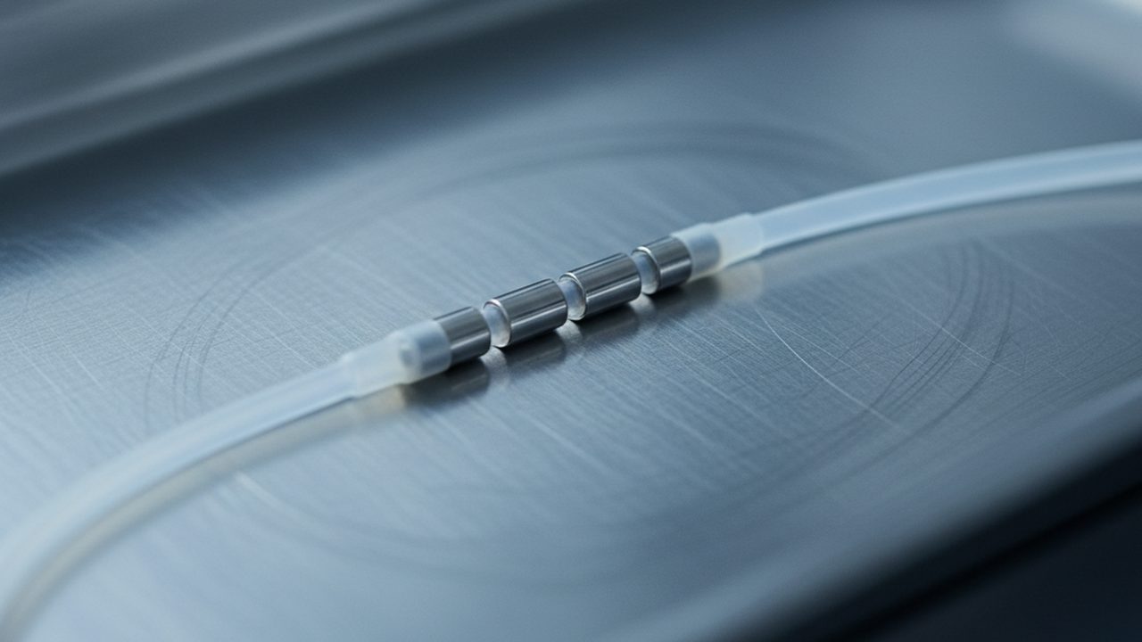
A DBS system consists of three primary components: the lead, the extension wire, and the implantable pulse generator. The lead is a thin wire, typically around 1.3 millimetres in diameter, which is guided through a small opening in the skull. At its tip are four or more cylindrical contacts made of a platinum-iridium alloy. These contacts are the only parts of the lead that are not insulated, allowing the electrical current to flow into the surrounding brain tissue.
The lead is connected to an extension wire that runs under the skin of the scalp and neck to the pulse generator, which is usually implanted beneath the collarbone. This generator contains the battery and the microcircuitry necessary to produce the electrical pulses. Modern generators are roughly the size of a large pocket watch and can be programmed wirelessly by a clinician. The settings include the voltage, the pulse width, and the frequency, which usually ranges between 130 and 185 Hertz for motor disorders.
The physical placement of the lead is a feat of stereotactic surgery. Because the target structures are often no larger than a grain of rice and are buried deep beneath the cortex, surgeons use a combination of magnetic resonance imaging and real-time microelectrode recording. During the procedure, the patient is often kept awake to provide feedback. As the electrode descends, the surgical team listens to the characteristic "signature" of different brain regions translated into sound. The subthalamic nucleus, for instance, has a distinct, rapid firing pattern that sounds like rain on a tin roof. Once the optimal site is identified, the permanent lead is anchored to the skull.
Mechanism of high-frequency inhibition in the basal ganglia
It was originally assumed that DBS worked by stimulating the target area to increase its output. However, clinical observations quickly suggested the opposite. The effects of high-frequency stimulation often mimicked the effects of a lesion, where the brain tissue is physically destroyed. If you destroy a hyperactive part of the basal ganglia, symptoms improve; if you apply high-frequency stimulation to that same spot, symptoms also improve. This led to the paradox that stimulation was, in a functional sense, acting as an inhibition.
The device functions as a high-frequency jammer, drowning out the brain's internal noise with a steady, predictable hum.
The prevailing theory for this mechanism involves the concept of informational masking. The high-frequency pulses likely drive the neurons at a rate so fast and so regular that they can no longer transmit their own pathological, low-frequency oscillations. In Parkinson’s disease, the basal ganglia often become stuck in a state of excessive synchrony, where neurons fire together in rhythmic bursts. This synchrony acts as a form of neural gridlock, preventing the brain from sending clear commands to the muscles. The DBS electrode introduces a constant signal that breaks this synchrony, effectively clearing the path for other, healthier circuits to function.
There is also evidence that DBS affects the white matter tracts, the "cables" of the brain, rather than just the grey matter cell bodies at the tip of the electrode. By stimulating these tracts, the device can influence distant parts of the brain that are connected to the target site. This means the mechanism is not local but architectural. It resets the rhythm of an entire network, allowing the cortex to regain control over motor output without the interference of the diseased deep-brain structures.
Target selection for motor and mood disorders
The success of DBS depends entirely on the selection of the target. For Parkinson’s disease, the most common targets are the subthalamic nucleus and the globus pallidus internus. Both are components of the basal ganglia that become overactive when dopamine is absent. Stimulating these areas reduces the inhibitory signal they send to the thalamus, which in turn allows the thalamus to properly excite the motor cortex. The result is a reduction in tremor and a restoration of the ability to initiate movement.
Targeting for mood disorders, such as treatment-resistant depression, is more complex and remains a subject of active research. One primary target is the subcallosal cingulate, an area of the brain that acts as a junction for circuits involving emotion, memory, and reward. In many depressed patients, this area shows chronic hyperactivity. Researchers have found that by applying stimulation here, they can "down-regulate" this activity, which often correlates with a lifting of the heavy, physical symptoms of depression.
Another promising target for psychiatric conditions is the ventral striatum and the internal capsule. These areas are involved in the brain's reward system and the processing of motivation. For patients with severe obsessive-compulsive disorder, stimulation in this region can disrupt the repetitive loops of thought that characterise the condition. Unlike motor targets, where the effect is nearly instantaneous, the effects of stimulation on mood can take weeks or months to fully manifest. This suggests that while the electrical intervention is immediate, the brain requires time to physically reconfigure its synaptic connections in response to the new rhythm.
The field is currently moving toward what is known as closed-loop stimulation. While traditional DBS provides a constant, unchanging pulse, closed-loop systems are designed to sense the brain's activity and only deliver a pulse when a specific pathological signal is detected. This approach aims to reduce side effects and prevent the brain from habituating to the stimulation. It represents the next step in the refinement of the interface, moving from a steady beat to a responsive dialogue with the brain's internal circuitry.
The limitations of open-loop continuous stimulation
Standard deep brain stimulation operates as an open-loop system. The pulse generator sends a continuous, rhythmic electrical current to the electrode at a fixed frequency and voltage, regardless of the patient's fluctuating state or current activity. While this has proved effective for controlling the tremors of Parkinson’s disease, it remains a relatively blunt instrument. The brain is not a static organ; its electrical requirements change throughout the day, during sleep, and as the underlying disease progresses.
Continuous stimulation carries several practical and clinical disadvantages. By flooding a specific nucleus with a constant signal, the system may inadvertently stimulate adjacent brain structures, leading to side effects such as speech impairment, mood changes, or motor contractions. In some patients, the brain appears to adapt to the constant signal, a process sometimes called tolerance, requiring clinicians to gradually increase the voltage to maintain the same therapeutic effect. This escalation often brings the patient closer to the threshold where side effects become intolerable.
Furthermore, a constant signal is inefficient. In many neurological conditions, the pathological brain activity that causes symptoms is not present every second of the day. In Parkinson’s disease, for example, the characteristic beta-frequency oscillations that interfere with movement are episodic. Providing stimulation during periods when the brain is functioning normally is unnecessary and may interfere with healthy neural processing. Open-loop systems also lack the ability to provide data back to the physician, meaning adjustments are typically made based on subjective clinical observation rather than objective measurements of brain state.
Sensing local field potentials for adaptive response
To move beyond these constraints, engineers have developed systems capable of sensing local field potentials, or LFPs. These are the aggregate electrical signals produced by thousands of neurons in the vicinity of the electrode tip. By listening to these signals, the device can identify specific biomarkers—distinctive patterns of electrical activity that correlate with particular symptoms. When the device detects the onset of a pathological rhythm, it triggers a pulse of stimulation to suppress it, then returns to a dormant sensing state once the rhythm dissipates.
The device functions less like a pacemaker and more like a thermostat, responding only when the internal environment shifts away from the desired range.
The primary biomarker currently used in research for Parkinson’s disease is the beta band, a signal between 13 and 30 hertz. High-intensity beta oscillations in the subthalamic nucleus are strongly associated with bradykinesia, or slowness of movement. In a closed-loop or adaptive system, the implant monitors the power of this beta signal in real time. If the power exceeds a predetermined threshold, the stimulation ramps up. As the patient moves and the beta signal naturally subsides, the stimulation decreases.
This sensing capability provides a window into the pathophysiology of the disease that was previously unavailable. It allows researchers to see how medication, sleep, and physical activity change the electrical signature of the brain over months or years. However, identifying reliable biomarkers for psychiatric conditions, such as depression or obsessive-compulsive disorder, remains significantly more difficult than for motor disorders. These conditions often involve widely distributed networks rather than a single focal point, and the electrical signature of a low mood is less distinct than the signature of a physical tremor.
Engineering constraints of battery life and lead migration
Transitioning from a passive stimulator to an active, sensing computer inside the skull introduces significant engineering challenges. The most immediate of these is power consumption. Sensing and processing neural data in real time requires more energy than simply firing a constant pulse. For a device that must remain implanted for years, battery life is a critical safety consideration. Current devices typically use primary cell batteries that last between three and five years, or rechargeable units that require the patient to wear a charging collar for several hours each week.
The physical stability of the hardware is another concern. The brain is a soft, gelatinous tissue, whereas the electrodes are relatively rigid. Although the leads are anchored to the skull, they are subject to micro-movements caused by the pulsing of blood vessels and the natural shifting of the brain within the cerebrospinal fluid. Over time, a lead may migrate by a few millimetres, moving the contact points away from the intended target. Even a small displacement can render the stimulation ineffective or cause new side effects by stimulating the wrong circuit.
There is also the issue of the foreign body response. When an electrode is inserted, the brain responds by forming a layer of glial scarring, or gliosis, around the hardware. This scar tissue is non-conductive and creates impedance, making it harder for the electrical signal to reach the target neurons. In a sensing system, this impedance can also muffle the local field potentials, making it harder for the device to "hear" the brain. Engineers must design the electrode surfaces and the stimulation waveforms to minimise this inflammatory response, often using materials like platinum-iridium and pulse shapes that balance the charge to prevent tissue damage.
Neuroplasticity and the long-term effects of chronic stimulation
While DBS was originally conceived as a way to "jam" or inhibit overactive circuits, it is now understood that chronic stimulation induces long-term changes in the brain's structure and function. This is particularly evident in the treatment of dystonia, a movement disorder where the benefits of stimulation often take weeks to appear and can persist for days or even weeks if the device is turned off. This suggests that the electrical pulses are not just masking symptoms, but are encouraging the brain to rewire itself through neuroplasticity.
Plasticity involves the strengthening or weakening of synaptic connections between neurons. By consistently disrupting pathological rhythms, DBS may allow healthier patterns of communication to re-emerge. There is emerging evidence that long-term stimulation can alter the expression of certain genes and the production of neurotrophic factors, which are proteins that support the growth and survival of neurons. In some cases, this leads to a stabilising effect where the patient requires less stimulation over time to achieve the same result.
However, the long-term effects of chronic electrical interference are not entirely understood. There is a theoretical risk that years of stimulation could lead to permanent changes in a patient's personality or cognitive abilities, particularly when the electrodes are placed near regions involved in emotion and executive function. Studies of long-term cohorts have generally shown that DBS is safe, but the variability in how individual brains adapt to the implant means that monitoring must be lifelong. The goal of future systems is to harness this plasticity, using the device to "train" the brain back into a healthy state until the stimulation is no longer required.
The move toward individualised circuit maps
The next frontier in deep brain stimulation is the transition from anatomical targeting to functional circuit mapping. Traditionally, neurosurgeons have targeted specific physical structures, such as the subthalamic nucleus, based on their appearance on an MRI scan. However, it is becoming clear that the success of the treatment depends less on stimulating a specific "blob" of gray matter and more on modulating the white matter tracts—the bundles of axons that connect different regions of the brain.
This approach, often called tractography-based targeting, uses advanced imaging techniques like diffusion tensor imaging to map the unique wiring of an individual patient's brain. Two patients may have electrodes in the exact same anatomical location, but because their internal wiring differs slightly, the stimulation may affect different downstream circuits. By mapping these connections before surgery, clinicians can tailor the placement of the electrode to intersect with the specific pathways responsible for the patient's symptoms.
- Anatomical targeting focuses on the location of the electrode tip within a specific nucleus.
- Circuit-based targeting focuses on the network of brain regions that the electrode can influence.
This shift is particularly important for expanding DBS into the treatment of complex conditions like Alzheimer’s disease or treatment-resistant depression. In these cases, there is no single "broken" part of the brain; instead, there is a malfunction in the way various regions communicate. The move toward individualised maps suggests a future where the implant is not just a standard medical device, but a bespoke intervention tuned to the unique geometry of an individual's neural architecture.
The current state of deep brain stimulation is one of established efficacy for motor disorders and promising, though still contested, results for psychiatric conditions. It is well-established that high-frequency stimulation can suppress tremors and improve quality of life for thousands of patients. What remains unresolved is the optimal way to sense and respond to the brain's internal state in real time, and whether the biomarkers we currently use are the most meaningful indicators of disease.
The field would be fundamentally changed by the discovery of a universal biomarker for mood or cognition, or by the development of wireless, leadless electrodes that could be implanted without the risks associated with long wires and bulky batteries. Until then, DBS remains the most sophisticated tool available for the direct modulation of the human nervous system, providing a functional bridge between the world of electronics and the delicate rhythms of the brain.